


This manual serves introductory human anatomy and physiology students‚ offering diverse lab experiences. It visualizes structures‚ links them to functions‚ and explores homeostasis.
Course Overview & Objectives
This anatomy lab course establishes a foundational knowledge of human anatomy‚ emphasizing the identification of anatomical structures through engaging‚ hands-on activities and relevant clinical applications. The curriculum adopts a regional approach‚ beginning with foundational concepts in histology‚ radiology‚ and a comprehensive overview of the body’s systems.
Subsequently‚ the course delves into detailed explorations of three distinct body regions: the thorax‚ abdomen‚ and pelvis. Students will gain practical skills in anatomical dissection and observation‚ fostering a deeper understanding of the interconnectedness of bodily structures and their functions.
Lab Safety Protocols
Prioritizing safety is paramount within the human anatomy and physiology laboratory. Students must adhere strictly to all outlined protocols to ensure a secure learning environment. This includes wearing appropriate personal protective equipment (PPE) – lab coats‚ gloves‚ and eye protection – at all times during dissection and experimentation.
Proper handling and disposal of specimens‚ chemicals‚ and sharp instruments are crucial. Familiarize yourself with emergency procedures‚ including the location of safety equipment like eyewash stations and first aid kits. Report any accidents or unsafe conditions immediately to the instructor. Maintaining a clean and organized workspace is also essential.

Cell Structure & Function
This section explores the foundational units of life‚ examining cellular components and their roles in maintaining physiological processes within the human body.
Microscope Use & Techniques
Effective microscopy is crucial for understanding human anatomy and physiology. This lab focuses on mastering techniques for optimal specimen visualization. Students will learn proper microscope handling‚ including focusing procedures‚ illumination adjustments‚ and objective lens selection.
Emphasis will be placed on preparing wet mounts and stained slides‚ essential for observing cellular structures. Techniques like focusing and depth of field will be practiced. Understanding magnification calculations and resolving power limitations are key. Proper cleaning and storage protocols will also be covered‚ ensuring longevity of this vital lab equipment.
Cellular Components: Organelles
This lab explores the intricate world within cells‚ focusing on key organelles and their functions. Students will identify and describe structures like the nucleus‚ mitochondria‚ ribosomes‚ endoplasmic reticulum‚ Golgi apparatus‚ and lysosomes. Microscopic observation of prepared slides will reveal these components‚ linking structure to specific cellular processes.
Understanding the role of each organelle in maintaining cellular life is paramount. Activities will emphasize how organelles collaborate to perform essential tasks like protein synthesis‚ energy production‚ and waste removal. This foundational knowledge is critical for comprehending tissue and organ-level physiology.
Cell Membrane Transport Mechanisms
This lab investigates how substances move across the cell membrane‚ a crucial aspect of cellular function. Students will explore passive transport – diffusion‚ osmosis‚ and facilitated diffusion – and active transport‚ requiring energy expenditure. Experiments will demonstrate the impact of concentration gradients‚ membrane permeability‚ and carrier proteins on transport rates.
Observing osmosis in different solutions and modeling active transport will solidify understanding. The importance of these mechanisms in maintaining cellular homeostasis‚ nutrient uptake‚ and waste removal will be emphasized. This knowledge forms a basis for understanding physiology at the tissue and organ levels.
Histology: The Study of Tissues
Histology labs focus on tissue types – epithelial‚ connective‚ muscle‚ and nervous – examining their structures and relating them to specific functions within the body.
Epithelial Tissue Types & Characteristics
Epithelial tissues cover body surfaces and line body cavities‚ exhibiting diverse types based on shape and layering. Students will investigate squamous‚ cuboidal‚ and columnar epithelia‚ noting their roles in protection‚ secretion‚ absorption‚ and filtration.
Labs emphasize identifying simple and stratified arrangements‚ alongside specialized features like cilia and microvilli. Understanding these characteristics is crucial for correlating structure with function across various organ systems. Practical exercises involve microscopic observation of prepared slides‚ fostering skills in histological identification and analysis. This foundational knowledge supports comprehension of tissue-level responses to physiological changes.
Connective Tissue: Classification & Functions
Connective tissues provide support‚ connection‚ and protection within the body‚ differing significantly from epithelial tissues. This lab explores classifications – connective tissue proper (loose & dense)‚ supporting cartilage‚ bone‚ and fluid connective tissues (blood).
Students will analyze the matrix composition and cellular components unique to each type. Emphasis is placed on relating structural features to specific functions like binding‚ transport‚ and immune defense. Microscopic examination of prepared slides will aid in identifying tissue characteristics‚ enhancing understanding of their roles in maintaining overall body homeostasis and structural integrity.
Muscle Tissue: Skeletal‚ Smooth‚ & Cardiac
Muscle tissues are specialized for contraction‚ enabling movement. This lab focuses on the three types: skeletal‚ smooth‚ and cardiac. Students will differentiate these tissues based on microscopic structure – striations‚ cell shape‚ and nucleus count.
Emphasis will be placed on correlating structural differences with functional roles. Skeletal muscle facilitates voluntary movement‚ smooth muscle controls involuntary processes in internal organs‚ and cardiac muscle powers the heart. Prepared slides will be examined to identify key features‚ solidifying understanding of muscle tissue function and its vital contribution to physiological processes.
Nervous Tissue: Neurons & Glial Cells
Nervous tissue forms the rapid communication network within the body. This lab explores its cellular components: neurons and glial cells. Students will identify neuron structures – cell body‚ dendrites‚ and axon – and understand their roles in signal transmission.
Microscopic observation of prepared slides will reveal different types of glial cells (astrocytes‚ oligodendrocytes‚ microglia) and their supportive functions. Emphasis will be placed on the neuron’s ability to generate and propagate electrical signals‚ and how glial cells contribute to neuronal health and efficient nervous system operation.

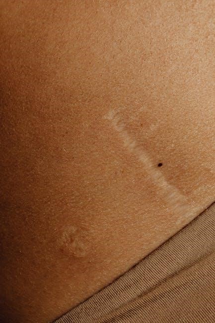
The Integumentary System
This lab investigates the skin – epidermis‚ dermis‚ and accessory structures like hair and glands – exploring their structure and vital protective functions.
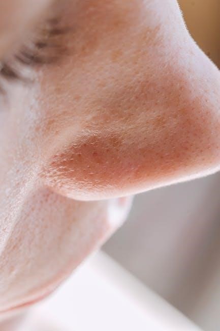
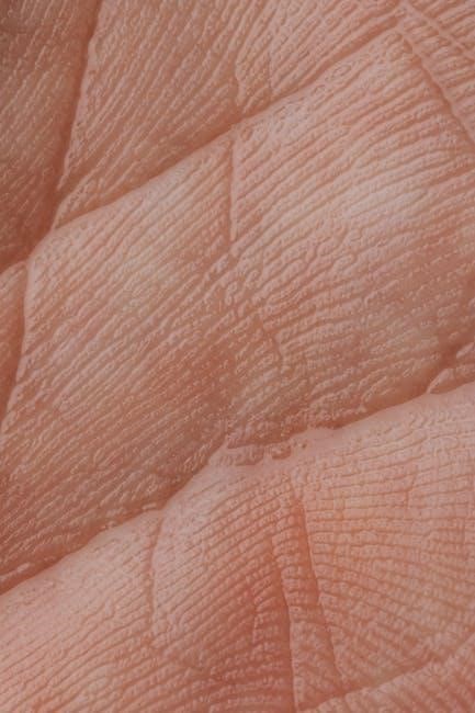
Epidermis: Layers & Functions
The epidermis‚ the outermost layer‚ is crucial for protection. Lab exercises focus on identifying its distinct layers – stratum corneum‚ lucidum‚ granulosum‚ spinosum‚ and basale – each with specialized cells. Students will examine keratinocyte migration and melanin production‚ understanding their roles in waterproofing and UV protection.
Activities explore the epidermis’s barrier function against pathogens and dehydration‚ alongside its sensory receptors for touch and temperature. The manual details how epidermal ridges contribute to grip and fingerprint formation‚ linking structure to physiological roles. Observing microscopic slides reveals cellular arrangements and layer thicknesses‚ solidifying comprehension of epidermal anatomy and function.
Dermis: Structure & Components
The dermis‚ lying beneath the epidermis‚ provides strength and elasticity. Lab investigations dissect its two layers: papillary and reticular. Students will identify collagen and elastin fibers responsible for skin’s resilience‚ alongside blood vessels nourishing the epidermis.
Exercises focus on sensory receptors – Meissner’s corpuscles‚ Pacinian corpuscles‚ and free nerve endings – relating structure to tactile perception. The manual details the role of dermal papillae in forming fingerprints and enhancing grip. Observing microscopic slides reveals fiber arrangements and component distribution‚ reinforcing understanding of dermal anatomy and its vital physiological functions.
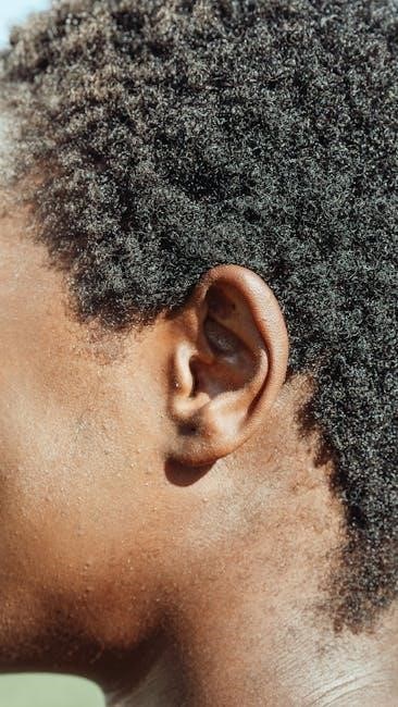
Accessory Structures: Hair‚ Nails‚ & Glands
This lab explores hair follicles‚ observing hair structure and growth phases through microscopy. Students analyze nail composition – keratin – and identify nail structures like the matrix and lunula. Investigations detail the function of sebaceous glands‚ relating their secretion to skin lubrication and acne formation.
Sweat gland types – eccrine and apocrine – are examined‚ linking their distribution to thermoregulation and scent production. Dissection exercises reveal the relationship between accessory structures and the dermis‚ emphasizing their roles in protection and homeostasis. The manual reinforces anatomical identification and physiological significance.
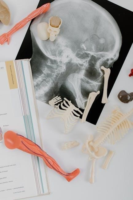
This lab introduces bone classification and structure‚ preparing students for skeletal system exploration. It covers axial and appendicular skeletons through practical exercises.
Bone Classification & Structure
This laboratory segment focuses on categorizing bones – long‚ short‚ flat‚ irregular‚ and sesamoid – based on their shapes and functions within the skeletal system. Students will delve into the microscopic and macroscopic structures of bone tissue‚ examining compact and spongy bone arrangements.
Detailed observation of bone components like osteons‚ Haversian canals‚ and Volkmann’s canals will be conducted. The composition of bone matrix‚ including organic and inorganic components‚ will be analyzed. Practical exercises will involve identifying different bone markings and features‚ understanding their roles in muscle attachment and joint articulation‚ ultimately solidifying comprehension of skeletal framework.
Axial Skeleton: Skull‚ Vertebral Column‚ & Rib Cage
This lab explores the axial skeleton’s components: the skull (cranial and facial bones)‚ vertebral column (cervical‚ thoracic‚ lumbar‚ sacral‚ and coccygeal vertebrae)‚ and the rib cage (true‚ false‚ and floating ribs). Students will identify individual bones‚ sutures‚ foramina‚ and processes.
Dissection and model analysis will highlight the protective functions of the skull and rib cage‚ alongside the vertebral column’s support and flexibility. Emphasis will be placed on understanding spinal curves and intervertebral discs. Practical application involves recognizing anatomical landmarks and their clinical relevance‚ fostering a comprehensive understanding of axial support.
Appendicular Skeleton: Limbs & Girdles
This lab focuses on the appendicular skeleton – the bones of the limbs and their supporting girdles (pectoral and pelvic). Students will identify bones of the upper limbs (humerus‚ radius‚ ulna‚ carpals‚ metacarpals‚ phalanges) and lower limbs (femur‚ tibia‚ fibula‚ tarsals‚ metatarsals‚ phalanges).
Practical exercises involve examining bone markings and articulating joints. The pectoral girdle (clavicle and scapula) and pelvic girdle will be analyzed for their role in limb attachment and movement. Emphasis is placed on understanding limb leverage and how bone structure supports locomotion and manipulation.
Joints: Articulations
This lab explores joint classifications – fibrous‚ cartilaginous‚ and synovial – and their structural characteristics. Students will analyze synovial joint components and movements.
Joint Classification: Fibrous‚ Cartilaginous‚ & Synovial
This laboratory section focuses on categorizing joints based on their structure and function. Fibrous joints‚ generally immovable‚ are connected by dense connective tissue. Cartilaginous joints allow limited movement‚ utilizing cartilage for connection‚ like intervertebral discs.
Synovial joints‚ the most common type‚ provide substantial movement‚ featuring a joint cavity filled with synovial fluid. Students will examine the unique characteristics of each classification‚ identifying examples within the skeletal system.
The lab emphasizes understanding how structural differences dictate the range and type of motion possible at each joint‚ linking anatomy directly to physiological function. Dissection and modeling activities will reinforce these concepts.
Synovial Joint Structure & Movements
This lab delves into the intricate anatomy of synovial joints – the most prevalent joint type enabling significant movement. Students will dissect and identify key components: articular cartilage‚ the joint capsule‚ synovial membrane‚ and ligaments.
Emphasis is placed on understanding how these structures contribute to joint stability and reduce friction. The lab explores various movements possible at synovial joints – flexion‚ extension‚ abduction‚ adduction‚ rotation‚ and circumduction.
Practical exercises involve identifying these movements on anatomical models and correlating them with specific muscle actions‚ solidifying the link between structure and function.
The Lower Limb
This lab investigates the lower limb‚ covering bones‚ muscles‚ joints‚ nerves‚ and blood vessels. Students will dissect and identify structures for functional understanding.
Bones of the Lower Limb
This laboratory focuses on identifying the bones comprising the lower limb‚ essential for understanding its structure and function. Students will examine the femur‚ tibia‚ fibula‚ tarsals‚ metatarsals‚ and phalanges.
Detailed observation and palpation exercises will reinforce anatomical knowledge. The lab emphasizes recognizing bony landmarks crucial for muscle attachment and joint articulation. Comparative analysis of bone shapes and sizes will highlight adaptations for weight-bearing and locomotion.
Furthermore‚ students will explore the skeletal framework supporting the lower limb‚ relating bone structure to its role in movement and stability. This foundational knowledge is vital for subsequent study of muscles and joints.
Muscles of the Lower Limb: Compartments
This lab investigates the muscles of the lower limb organized into distinct compartments – anterior‚ posterior‚ medial‚ and lateral – each containing muscles with shared innervation and function. Students will identify key muscles within each compartment‚ noting their origins‚ insertions‚ and actions.
Dissection and palpation exercises will enhance understanding of muscle location and relationships. Emphasis will be placed on correlating compartment organization with specific movements like dorsiflexion‚ plantarflexion‚ inversion‚ and eversion.
Analyzing muscle arrangements reveals how compartments contribute to efficient and coordinated lower limb movement.
Joints of the Lower Limb: Hip‚ Knee‚ Ankle
This lab focuses on the structural classifications and functional characteristics of the hip‚ knee‚ and ankle joints. Students will examine the hip as a ball-and-socket joint‚ the knee as a modified hinge joint‚ and the ankle as a complex joint enabling dorsiflexion and plantarflexion.
Dissection will reveal ligamentous support and meniscal structures within each joint. Range-of-motion exercises will demonstrate joint movements and stability.
Understanding joint mechanics is crucial for comprehending lower limb function and potential injury mechanisms.
Nerves & Blood Vessels of the Lower Limb
This lab investigates the intricate network of nerves and blood vessels supplying the lower limb. Students will trace the pathways of major arteries – femoral‚ popliteal‚ and anterior/posterior tibial – and veins‚ understanding their branching patterns and collateral circulation.
Dissection will reveal the course of key nerves‚ including the femoral‚ obturator‚ and sciatic nerves‚ and their major branches.
Emphasis will be placed on correlating neurovascular structures with muscle innervation and blood supply‚ crucial for clinical applications.
The Upper Limb
This lab focuses on the upper limb’s anatomy‚ dissecting bones‚ muscles‚ and joints. Students will identify compartments and explore nerve/vessel pathways.
Bones of the Upper Limb
This laboratory investigates the skeletal framework of the upper limb‚ beginning with the clavicle and scapula‚ forming the shoulder girdle. Students will meticulously identify the humerus‚ radius‚ and ulna – the bones comprising the arm and forearm.
Detailed examination includes recognizing key landmarks‚ articulating surfaces‚ and bony projections crucial for muscle attachment and joint articulation. Practical exercises involve assembling the skeletal components to understand their spatial relationships and functional integration.
Emphasis is placed on differentiating between the bones and understanding their contribution to the overall range of motion and stability of the upper limb.
Muscles of the Upper Limb: Compartments
This lab focuses on the organization of muscles within the upper limb‚ categorized into distinct compartments – anterior and posterior – of both the arm and forearm. Students will identify and locate muscles within each compartment‚ noting their origins‚ insertions‚ and primary actions.
Dissection and palpation exercises enhance understanding of muscle relationships and functional groupings. Emphasis is placed on recognizing how compartment boundaries influence muscle function and nerve/vessel pathways.
Students will correlate muscle compartment organization with specific movements of the shoulder‚ elbow‚ wrist‚ and hand.
Joints of the Upper Limb: Shoulder‚ Elbow‚ Wrist
This lab investigates the structural classifications and functional characteristics of the upper limb’s major joints: the shoulder (glenohumeral)‚ elbow (humeroulnar & humeroradial)‚ and wrist (radiocarpal). Students will dissect and analyze these joints‚ identifying key ligaments‚ articular surfaces‚ and associated bursae.
Range of motion exercises demonstrate joint capabilities‚ while correlating movements with muscle actions reinforces understanding. Emphasis is placed on joint stability and potential injury mechanisms.
Students will explore how joint structure dictates specific movements and contributes to overall upper limb functionality.
Nerves & Blood Vessels of the Upper Limb
This lab focuses on identifying the major nerves and blood vessels supplying the upper limb‚ tracing their pathways from origin to distribution. Dissection reveals the brachial plexus and its primary branches – musculocutaneous‚ median‚ ulnar‚ and radial nerves – alongside associated arteries and veins.
Students will correlate nerve innervation with specific muscle groups and assess vascular supply to different regions of the arm‚ forearm‚ and hand.
Understanding neurovascular relationships is crucial for diagnosing and treating upper limb injuries.

Laboratory Techniques & Dissection
Labs emphasize proper dissection procedures‚ anatomical terminology‚ and directional terms. Students learn specimen preservation‚ handling‚ and accurate identification of structures;
Proper Dissection Procedures
Successful dissection requires careful planning and adherence to established protocols. Begin with a thorough understanding of the specimen’s anatomy and the specific structures to be identified. Utilize appropriate dissection tools – scalpels‚ scissors‚ probes – with precision and caution‚ always cutting away from yourself and others.
Maintain a clean and organized workspace‚ and properly dispose of biological waste according to laboratory guidelines. Gentle teasing apart of tissues is often preferable to forceful cutting‚ preserving delicate structures. Accurate observation and documentation‚ including sketches and labeling‚ are crucial for learning. Respectful handling of the specimen is paramount‚ recognizing its origin and purpose in anatomical study.
Anatomical Terminology & Directional Terms
Mastering anatomical language is fundamental to understanding human anatomy. Standardized terminology ensures clear communication‚ avoiding ambiguity when describing locations and relationships of body structures. Directional terms – superior‚ inferior‚ anterior‚ posterior‚ medial‚ lateral‚ proximal‚ distal – establish a framework for precise anatomical descriptions.
Planes of section – sagittal‚ frontal (coronal)‚ transverse – define how the body is viewed. Regional terms categorize body areas‚ while root words‚ prefixes‚ and suffixes build a comprehensive anatomical vocabulary. Consistent use of this terminology is essential for accurate observation‚ dissection‚ and clinical application.
Specimen Preservation & Handling
Proper specimen handling is crucial for maintaining anatomical integrity and ensuring safe laboratory practices. Preservatives‚ like formalin‚ prevent decomposition but require careful handling due to toxicity. Always wear appropriate personal protective equipment – gloves‚ goggles‚ and lab coats – when working with preserved specimens.
Gentle handling minimizes tissue damage. Return specimens to designated containers after use‚ and follow disposal protocols. Understanding preservation techniques and safety guidelines is paramount for effective learning and responsible laboratory conduct‚ respecting the material and ensuring a safe environment.
Homeostasis & Physiological Processes
Labs explore homeostasis‚ examining feedback mechanisms – negative and positive – alongside foundational concepts in cardiovascular and respiratory physiology.
Feedback Mechanisms: Negative & Positive
Understanding how the body maintains stability is crucial. This section delves into feedback mechanisms‚ the core of homeostasis. Negative feedback‚ the more common type‚ reverses a change to maintain a set point – think body temperature regulation. Conversely‚ positive feedback amplifies a change‚ often seen in blood clotting or childbirth.
Laboratory exercises will likely involve scenarios demonstrating these principles‚ perhaps simulating blood glucose control or observing the cascade of events in coagulation. Students will analyze data and predict responses to disruptions‚ solidifying their grasp of these vital physiological processes and their importance in overall health.
This section introduces the fascinating world of the cardiovascular system – the heart‚ blood vessels‚ and blood. Labs will focus on understanding the heart’s anatomy‚ its conduction system‚ and the mechanics of cardiac output. Students will likely measure heart rate and blood pressure‚ exploring factors that influence these vital signs.
Experiments may involve analyzing ECGs‚ observing blood flow dynamics‚ and investigating the role of different blood components. The goal is to grasp how this system delivers oxygen and nutrients while removing waste‚ maintaining homeostasis and supporting all bodily functions.
This module delves into the mechanics of breathing and gas exchange within the human respiratory system. Laboratory exercises will likely involve measuring lung volumes using spirometers‚ analyzing breathing rates‚ and exploring the impact of exercise on respiratory function. Students will investigate the anatomy of the lungs and airways‚ understanding how structure relates to function.
Experiments may also examine gas diffusion across the alveolar-capillary membrane and the role of hemoglobin in oxygen transport. The aim is to comprehend how the respiratory system collaborates with the cardiovascular system to maintain homeostasis and deliver oxygen to tissues.
Leave a Reply
You must be logged in to post a comment.